Fatty Acid Pathways and Lipid Metabolism in HER2+ Breast Cancer
How HER2 signaling reprograms fat-building, fat-burning, and cholesterol pathways in HER2-positive breast cancer, and what this means for treatment resistance and targeted drugs.
The same HER2 → PI3K/AKT/mTOR signaling described above for glucose also reprograms how HER2+ cells build, store, and use fat. These changes are not a set of separate quirks — they form a single connected assembly line, where the output of one enzyme becomes the raw material for the next, and HER2 signaling switches on several points along that line at once.
Contents Menu
This page works through six connected mechanisms, one at a time:
ACLY/PDH — converts leftover sugar-processing byproducts into the raw material used to build new fat
ACC — commits that raw material to fat production
SREBP-1/FASN — the master switch and finishing step for building new fat
FAO/CD36 — burning fat for fuel, and importing fat from outside the cell
SREBP-2/Mevalonate — the cholesterol-building branch of the same system
Macropinocytosis — a separate, non-manufacturing route that scavenges cholesterol directly from outside the cell, rather than building it
A fully referenced summary is available for integrative cancer practitioners and patients who want a deeper understanding. [insert link]
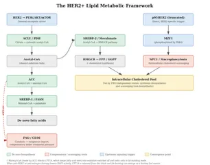
The HER2+ lipid metabolic framework: how fat-building, fat-burning, and cholesterol pathways connect. [insert better flow diagram]
ACLY/PDH: Turning Leftover Sugar into Fat
Before a HER2+ cancer cell can build new fat, it needs raw material — and that raw material typically starts as leftover sugar. Two connected enzymes control this handoff from sugar-burning to fat-building.
ACLY (ATP citrate lyase) takes citrate — a molecule made in the mitochondria from sugar-derived pyruvate — and converts it into acetyl-CoA, the basic building block cells use to make new fat and cholesterol.
PDH (pyruvate dehydrogenase) normally lets pyruvate get fully burned for energy inside the mitochondria. In HER2+ cells running the sugar-heavy metabolism described above, PDH gets partly switched off, which pushes pyruvate toward the citrate-export route instead — feeding the ACLY step.
These two enzymes function as two ends of the same shift: PDH suppression makes sure pyruvate is available for citrate export, and ACLY turns that exported citrate into fat-building material. Addressing one without the other may leave half the mechanism untouched.
HER2 signaling directly and specifically switches on ACLY. The chain runs: HER2 → PI3K → a particular branch of mTOR called mTORC2 → a chemical tag added to ACLY → more acetyl-CoA production.
This dependency has been shown in both ER-positive and ER-negative HER2+ lab models. It requires HER2 amplification together with a common companion mutation (in a gene called PIK3CA) — not ER status on its own.
PIK3CA mutations occur more often in triple-positive (ER+/HER2+) tumors than in ER-negative HER2+ tumors. So while the mechanism itself is not ER-driven, triple-positive tumors may be more likely to carry the specific mutation combination that switches it on.
In triple-positive disease specifically, estrogen signaling separately increases FASN — the enzyme that finishes fat production further down this same pathway (covered later in this section). HER2 and estrogen may therefore be pushing on two different points of the same assembly line at the same time.
Why this matters for HER2-targeted drugs
This pathway runs specifically through mTORC2, not mTORC1.
That distinction has a practical implication. Existing mTOR-blocking drugs used in HER2+ breast cancer, such as everolimus or temsirolimus, work mainly through mTORC1. On their own, they would not be expected to switch off this particular fat-building route. Blocking it pharmacologically would require a drug capable of hitting both mTORC1 and mTORC2 together.
A newly approved drug shows what that looks like in practice. Gedatolisib, which blocks all four types of PI3K alongside both mTORC1 and mTORC2, received FDA approval in July 2026 for hormone-receptor-positive, HER2-negative advanced breast cancer without a PIK3CA mutation. A second approval covering PIK3CA-mutant disease — the specific genetic combination this ACLY mechanism depends on — is expected later in 2026, based on trial results showing a substantial progression-free survival improvement over the prior standard treatment in that population.
Gedatolisib has not been tested in HER2+ or triple-positive breast cancer. New targeted drugs are typically tested first in ER+/HER2-negative disease before any HER2+ combination trial begins, and that next step, historically, tends to take years rather than months.
What we don't know yet
The mTORC2–ACLY connection comes from four cell lines (spanning both ER-positive and ER-negative HER2+ models), with no confirmation yet in a living HER2+ tumor model or in patient tissue.
Whether triple-positive tumors truly show more ACLY activity in practice remains an inference from mutation-frequency patterns, not a direct measurement in triple-positive tumor samples.
A further connection has been proposed but not tested: the same mTORC2 signaling that activates ACLY has separately been shown, in other research, to also stabilize SREBP-1 — the master switch for fat production covered in the next section. If both effects occur together in the same HER2+ tumor cell, mTORC2 could be coordinating fat production at two points in the pathway at once. This has not been directly tested as a single, unified mechanism in HER2+ breast cancer — it's a hypothesis worth tracking, not an established finding.
Whether the recently approved drug for HER+HER2- breast cancer called gedatolisib, or a similar drug that blocks PI3K and both mTOR complexes together, would affect this ACLY-driven route in HER2+/PIK3CA-mutant disease specifically has not been tested. It's a reasonable direction to watch, not a demonstrated benefit.
Sources: Chen et al., 2016; Hatzivassiliou et al., 2005; Li et al., 2016; Hurvitz et al., 2026
Most promising potential blocker for this pathway
METABLOC (hydroxycitric acid + R-ALA) (OTC)
ACC: Committing to Fat Production
Once ACLY has made acetyl-CoA, the next enzyme in the sequence makes the commitment. Acetyl-CoA carboxylase (ACC) converts acetyl-CoA into malonyl-CoA — the first step in this pathway that can't easily be reversed, and the actual starting material the next enzyme, FASN, uses to build new fat.
Cells make two versions of this enzyme with different jobs:
ACC1 sits in the cell fluid and drives fat production.
ACC2 sits at the mitochondrial membrane and works more like a brake on fat-burning: the malonyl-CoA it makes blocks CPT1A, the gatekeeper that lets fat into the mitochondria to be burned for fuel — the same CPT1A relationship shown in the diagram above.
Despite how extensively ACC has been studied in cancer generally, no dedicated study measuring ACC activity specifically in HER2+ breast cancer was identified in this review. That's a genuine, notable gap — not a case where HER2+ evidence points one way and other findings point another, but an area where HER2+-specific evidence simply doesn't yet exist.
ACC activity is switched off by an energy-sensing enzyme called AMPK. In one non-breast resistance study — head and neck cancer cells resistant to the antibody drug cetuximab — this AMPK-driven ACC shutdown initially worked as expected, but resistant cells compensated by making more total ACC protein, rewiring their metabolism from sugar-dependence toward fat-dependence despite the ongoing AMPK pressure. This kind of compensatory rebound, where blocking an enzyme's activity simply pushes cells to make more of it, is a pattern that comes up again later in this section, in a different part of the pathway.
In laboratory studies of breast cancer cells generally (not HER2+ specifically), turning off ACC or FASN triggered cell death, oxidative stress, and mitochondrial damage — effects that could be reversed by supplying the cells with palmitate (the fat these enzymes would otherwise have produced) or vitamin E, and that did not occur in normal, non-cancerous breast cells.
A related story: resistance to hormone-blocking drugs
Breast cancer cells that develop resistance to long-term estrogen deprivation — the same pressure created by aromatase inhibitors — develop an enhanced capacity to store fat, controlled specifically by ACC1. This stored-fat system is functionally linked to a separate cell structure that manages oxidative stress. Blocking ACC1 specifically impaired the survival of these resistant cells. This was confirmed not just in lab-grown cells, but in tissue samples and lab models built directly from patients previously treated with aromatase inhibitors.
This is directly relevant context for triple-positive patients on hormone-blocking therapy, since long-term aromatase-inhibitor use creates the same estrogen-deprivation pressure studied here. But it has not been tested in HER2+ or triple-positive tumors specifically — the underlying research was done in ER+ models without HER2 overexpression. Whether HER2 signaling changes this dynamic, in either direction, remains an open, untested question.
Separately, in one lab test, combining an ACC-blocking approach with a FASN-blocking approach (the next enzyme in this pathway, covered next) did not produce a bigger effect than blocking either one alone in hormone-therapy-resistant ER+ cells. That's a useful caution against assuming that targeting multiple connected steps in this pathway automatically adds up to a bigger benefit.
What we don't know yet
No study identified in this review has measured ACC activity directly in HER2+ breast cancer tissue or cell lines — a genuine gap in a pathway that is otherwise heavily studied in cancer generally.
The ACC1/fat-storage finding in hormone-therapy-resistant cells is a mechanistically plausible explanation for what might be happening in triple-positive patients on long-term aromatase-inhibitor therapy, but this remains an inference. It has not been directly tested in HER2+ or triple-positive disease.
Sources: Fullerton et al., 2013; Luo et al., 2017; Chajès et al., 2006; Foldi et al., 2022; Bacci et al., 2024; Balinda et al., 2025
Most promising potential blocker for this pathway
Metformin (Rx)
SREBP-1/FASN: The Switch and the Finishing Step
Once ACC has made malonyl-CoA, one more step finishes the job: turning that material into an actual usable fat molecule. Two things control this — a master switch that decides whether the whole system runs, and the enzyme that does the physical building.
SREBP-1 is the master switch. It sits inactive until growth signals and available nutrients tell the cell it's safe to build new fat, then it moves into the nucleus and turns on both ACC (the previous step) and FASN (the next one).
FASN (fatty acid synthase) does the actual construction, assembling malonyl-CoA units into palmitate — the finished fat molecule.
SREBP-1 gets switched on the same way SREBP-2 does elsewhere in this pathway: oncogenic PI3K signaling — the dominant signal coming out of HER2 — activates a branch of mTOR called mTORC1, which turns on SREBP-1. A second, separate route runs through mTORC2: losing mTORC2 signaling causes SREBP-1 to be broken down, which lowers both ACC and FASN activity.
This is the same mTORC2 branch covered earlier in this guide for ACLY. If both effects are real in the same HER2+ cell, mTORC2 could be controlling fat production at two points in the pathway at once — directly feeding the first step (ACLY) and indirectly keeping this master switch turned on. This remains an untested hypothesis bridging two separate studies, not an established mechanism.
The HER2-FASN feedback loop
HER2 and FASN reinforce each other in a loop: HER2 directly switches FASN on, and FASN activity, in turn, helps sustain HER2 signaling. The HER2-activating step is blocked by lapatinib; whether the newer HER2 drugs tucatinib and neratinib block this same step hasn't been directly tested but is probable.
In one lab study of actual HER2+ breast cancer cells that had become resistant to lapatinib, FASN was one of the clearest markers separating resistant cells from treatment-sensitive ones — a real, if single, HER2+-specific signal linking FASN to this particular kind of drug resistance.
Why this matters for HER2-targeted drugs
FASN is commonly elevated in HER2+ tumors, and tumors with both high HER2 and high FASN have worse outcomes than HER2+ tumors with lower FASN. Among ER+ tumors specifically, triple-positive tumors carry the highest FASN levels — higher than ER+/HER2-negative or basal-like disease.
Combining an mTOR-blocking drug with a FASN-blocking approach produced a notably stronger effect specifically in ER+/HER2+ models — not in models that were only ER-positive or only HER2-positive. This is one of the few findings in this whole framework that points specifically at triple-positive biology rather than at HER2+ or ER+ disease generally.
In ER+ disease more broadly (without HER2 involvement), FASN activity tends to stay elevated through resistance to several types of hormone-blocking treatment. But blocking FASN doesn't always translate into slower growth, even when it successfully reduces the fat being stored — a caution against assuming that hitting this enzyme automatically works, even in settings where it's clearly overactive. This particular finding comes from ER+/HER2-negative models only and hasn't been tested in triple-positive disease specifically.
No FASN-blocking drug is currently FDA-approved for any cancer.
What we don't know yet
Whether tucatinib and neratinib block the same HER2-to-FASN activation step lapatinib blocks remains untested.
The FASN/lapatinib-resistance finding above comes from a single study and needs replication before it can be treated as established.
The mTORC2 dual-role hypothesis described above — coordinating both ACLY and SREBP-1 at once — has not been tested as a single, unified mechanism in any HER2+ model.
Sources: Ricoult et al., 2016; Li et al., 2016; Jin et al., 2010; Vazquez-Martin et al., 2008; Steggall et al., 2025; Corominas-Faja et al., 2017; Menendez et al., 2021; Yan et al., 2014; Ward et al., 2025
A mechanistic counterpoint: palmitate itself
In lab studies, exposing HER2+ breast cancer cells directly to palmitate — the fat molecule FASN produces — killed those cells selectively, reduced HER2 and a related receptor (HER3), and made the cells more sensitive to trastuzumab. This is a single finding from three cell lines, has not been tested in animals or patient tissue, and should not be read as dietary guidance. It says something about how these particular cancer cells respond to a specific molecule in a dish — not about what to eat.
Sources: Baumann et al., 2016; Castagnoli et al., 2023
Most promising potential blocker for this pathway
EGCG / green tea extract (OTC)
FAO/CD36: Burning Fat, and Importing It From Outside the Cell
Fatty acid oxidation (FAO) is the mirror image of the fat-building steps just covered — instead of assembling new fat, mitochondria break existing fat down for fuel. The overview diagram [link to diagram] showed the switch that connects the two: when FASN is highly active, a byproduct called malonyl-CoA builds up and blocks CPT1A, the gatekeeper enzyme that lets fat into the mitochondria to be burned. That keeps the cell locked in fat-building mode. When anti-HER2 or anti-estrogen treatment lowers FASN activity, malonyl-CoA drops, CPT1A is released from that block, and fat-burning can switch on as an alternative fuel source.
Before fat can be burned, it also has to get into the cell in the first place:
CD36 imports long-chain fatty acids from outside the cell.
CPT1A then moves that fat into the mitochondria, where it's broken down for energy.
Together, CD36 and CPT1A form a two-step import-and-burn axis. A research group in Milan has built a coherent case that this whole axis functions as a HER2+ treatment-resistance mechanism.
CD36: the import step
In tissue from 180 patients in the NeoALTTO trial (validated in 331 more from the NeoSphere trial), higher CD36 levels in the tumor were independently linked to worse event-free survival — but only in patients treated with Herceptin alone, not in patients who received lapatinib or the trastuzumab-lapatinib combination. Patients with both high CD36 and no complete response to treatment had the worst outcomes of anyone in the study: a 7-year event-free survival of 47%, versus 79% for everyone else (Ligorio et al., 2022).
The researchers offered a theory for why this pattern showed up only with Herceptin: Herceptin partly relies on the immune system to kill cancer cells, while lapatinib doesn't. They thought CD36-high tumors might specifically blunt that immune-dependent mechanism. When they tested this directly — checking whether CD36 levels correlated with immune cell activity in tumors — they found no correlation. Their own proposed explanation wasn't supported by their own data, and they describe the question as still open.
Separately, CD36 was shown to increase specifically after anti-HER2 treatment, confirmed in two independent patient sample sets. This increase was concentrated in a particular subset of cancer stem-like cells, and biopsies from 36 HER2+ patients confirmed these CD36-high stem-like cells showed up significantly more often in patients who didn't achieve a complete response to treatment (Castagnoli et al., 2025).
A note on cancer stem cells and plant compounds
Cancer stem-like cells — the resistant, treatment-evading subpopulation described above — are an active area of botanical research generally. A range of plant-derived compounds have shown activity against cancer stem cell populations in laboratory studies, and combining this kind of activity with HER2-targeted therapy is an interesting research direction.
That said, "active against cancer stem cells" isn't one mechanism — different compounds work through very different routes. What's needed is identifying candidates that specifically target the CD36-high, Wnt-driven population, then checking for interactions with HER2-targeted drugs, tamoxifen, and taxane chemotherapy, since several compounds in this space affect the same liver enzyme (CYP3A4) that metabolizes those drugs.
CPT1A: the mitochondrial gatekeeper
In tissue from 430 breast cancer patients (not broken out by subtype), higher CPT1A levels tracked closely with disease progression, stage, and grade (Tan et al., 2021).
In HER2+ mouse models, researchers genetically engineered mice to lack a working copy of the CPT1A gene in their tumor cells — not a drug or supplement effect, but a research tool used to test what happens when this fat-burning gatekeeper is completely disabled. Tumors without CPT1A took longer to start growing, grew more slowly, and spread to the lungs less. Without CPT1A, the cells compensated by leaning harder on sugar for fuel — the same glycolytic dependence covered in the glucose section above — and by switching on a stress-response pathway called Nrf2.
Genetically removing CPT1A also helped the immune system attack the tumor more effectively, and combining CPT1A removal with an anti-HER2 antibody worked better than either approach alone. The study's own explanation for this combination benefit centers on the immune system: disabling CPT1A appears to shift the tumor's surroundings into a state more hospitable to immune attack, which then makes the anti-HER2 antibody's immune-dependent killing mechanism work better (Nandi et al., 2024).
There's a second, related idea worth flagging, though it isn't the explanation this specific study tested. The glucose section of this guide covers separate evidence that HER2-targeted antibodies like trastuzumab already reduce glucose uptake in HER2+ cells. If CPT1A-deficient cells become more dependent on glucose to survive, as this study found, a HER2-targeted drug that's simultaneously limiting glucose availability could be squeezing a cell that has just lost its fat-burning fallback option from a second direction at the same time. This connects two separately established findings — it isn't something Nandi et al. tested directly — and should not be read as an established mechanism.
A conference abstract (full paper not yet published) reported that genes involved in fat-burning were among the most active pathways in HER2+ tissue taken after Herceptin treatment, and that CPT1A activity was inversely linked to how well cells responded to lapatinib — meaning cells leaning more heavily on fat-burning tended to resist treatment more (Franceschini et al., 2023).
Why this matters for HER2-targeted drugs
A protein called PPARgamma appears to help drive this whole reprogramming — both the fat-building and fat-burning sides — in cells that have become resistant to anti-HER2 treatment. Blocking the PPARgamma pathway in lab studies restored anti-HER2 drug sensitivity in resistant cells. This connection is specific to HER2+ disease: the same pathway also helps maintain a cancer stem-cell population uniquely in HER2+ cells, with no equivalent effect seen in non-HER2+ cells tested side by side (Xiong et al., 2025; Wang et al., 2013).
What we don't know yet
No CPT1A-blocking drug has entered a clinical trial in HER2+ or triple-positive breast cancer, and no CD36-blocking drug is currently in trials for breast cancer at all.
The Franceschini finding above comes from a conference abstract with 33 paired tissue samples; no full peer-reviewed publication has been identified.
No study has directly compared fat-burning activity between triple-positive and ER-negative/HER2+ tumors.
Sources: Ligorio et al., 2022; Castagnoli et al., 2025; Tan et al., 2021; Nandi et al., 2024; Franceschini et al., 2023; Xiong et al., 2025; Wang et al., 2013
A related story: the same fuel-switch in ER+ disease, and specifically in triple-positive tumors
Fat-burning as a resistance mechanism isn't unique to HER2+ disease. In ER+HER2- breast cancer, a growing body of evidence links fat-burning to resistance against several types of hormone-blocking treatment, through more than one signaling route (Ahn et al., 2024; Jiang et al., 2023; Duan et al., 2021; Li et al., 2025).
In cell models built specifically from triple-positive breast cancer, developing resistance to aromatase-inhibitor therapy came with a rise in fat metabolism genes alongside a separate protective system that helps cells resist a specific kind of cell death driven by fat oxidation byproducts. Combining a drug that blocks this protective system with anti-HER2 treatment caused substantial cell death in these resistant models (Bahnassy et al., 2023).
No study has directly compared fat-burning activity between triple-positive tumors and ER-HER2+ tumors. But the existing evidence suggests triple-positive disease may face two separate pressures toward this fuel-switch at once: the anti-HER2 treatment resistance route (shared with ER-HER2+ disease) and a separate hormone-therapy resistance route specific to ER+ disease.
Sources: Ahn et al., 2024; Jiang et al., 2023; Duan et al., 2021; Li et al., 2025; Bahnassy et al., 2023
Ketogenic diet: what the evidence actually shows
In the same HER2+ mouse study described above, researchers also tested combining CPT1A loss with a ketogenic diet — a diet built around long-chain fatty acids, the exact fuel this pathway burns. That combination significantly slowed tumor growth beyond CPT1A loss alone, similar to what happened when CPT1A loss was combined with anti-HER2 antibody treatment instead (Nandi et al., 2024).
This result is easy to misread in isolation. It doesn't mean "ketogenic diet helps." When considered alongside everything above, it means something more specific: increasing the amount of fat available to a tumor, without also blocking the machinery (CPT1A) that lets the tumor use that fat, may hand the tumor more fuel rather than starve it. In this study, the diet only helped when paired with genetic CPT1A blockade — it was never tested as a standalone approach, and it has not been tested in humans at all.
This reframes the practical question. It isn't "should I try a ketogenic diet" — it's whether a ketogenic diet, if used, makes more mechanistic sense paired with something that also blocks fat-burning machinery, rather than used on its own in HER2+ disease. The current evidence — one mouse study — can't answer that directly, but the underlying mechanism is a real reason to ask the question before assuming a ketogenic diet is straightforwardly helpful here.
Sources: Nandi et al., 2024
Most promising potential blockers for this pathway
Ranolazine (Rx)
Omega-3 DHA / fish or algal oil (OTC)
SREBP-2/Mevalonate: The Cholesterol-Building Branch
The same acetyl-CoA that feeds fat production also feeds a second master switch: SREBP-2. Where SREBP-1 (covered above) turns on fat-building genes, SREBP-2 turns on cholesterol-building genes — and, just as importantly, genes that build two related molecules cells use to anchor certain growth-signaling proteins onto their inner membrane.
In HER2+ cells that have become resistant to HER2-targeted treatment, it's this anchoring function — not cholesterol itself — that seems to matter most for helping the cell survive.
Blocking this pathway runs into a specific problem. Statins (and supplement equivalents that work the same way, like citrus bergamot and red yeast rice) block the first, rate-limiting enzyme in this chain. But when the HER2+ cell senses the resulting drop in cholesterol, it switches SREBP-2 back on to rebuild everything just blocked — including the anchoring molecules the statin was trying to cut off. This rebound is a real limitation of statins used alone.
Two other approaches sidestep this problem, each in a different way:
Bisphosphonates (like zoledronic acid) block a step further downstream, past the point where the rebound kicks in — so they aren't subject to it.
Direct SREBP-2 blockers (dipyridamole, or the over-the-counter alternative gamma-tocotrienol) shut down the master switch itself, preventing the rebound from happening in the first place, rather than trying to out-block it.
Putting it into practice
Situation | Approach |
|---|---|
Not on Enhertu and taking (or considering) a statin, citrus bergamot, or red yeast rice | Pair it with an SREBP-2 blocker — dipyridamole (prescription) or gamma-tocotrienol (over-the-counter) |
Not on Enhertu and want to act on this pathway without a statin | Gamma-tocotrienol alone has its own direct SREBP-2-blocking mechanism. Dipyridamole doesn't work this way — its evidence is specifically about rescuing a statin, not standalone activity |
Not on Enhertu and already on a bisphosphonate | No pairing needed — the rebound problem doesn't apply to this drug class |
On Enhertu | Take a statin alone, without an SREBP-2 blocker. The rationale here is different (below), and adding dipyridamole adds a bleeding-risk concern on top of Enhertu's own risk, for no clear benefit |
Why a statin alone can rebound in HER2+ with HER2-targeted drugs (except Enhertu). [insert diagram]
Why this matters for HER2-targeted drugs
In the largest and most methodologically rigorous look at this question on today's standard HER2+ treatment backbone (Herceptin + Perjeta, or Phesgo), taking a statin on its own showed no survival benefit (Maurer et al., 2025). That's consistent with the rebound problem above — a statin alone, unpaired, may not be doing much in HER2+ breast cancer on most targeted treatments.
With Enhertu specifically, the logic flips. The goal isn't blocking the anchoring-molecule pathway — it's using cholesterol depletion to reduce how much HER2 gets pulled back inside the cell, leaving more HER2 on the surface for Enhertu to bind and deliver its payload. This membrane-level effect works even with a partial SREBP-2 rebound happening inside the cell, since the two processes run on different timescales. A small retrospective study in HER2-positive patients on Enhertu found statin use linked to meaningfully longer progression-free and overall survival, alongside supporting lab evidence (Sahin et al., 2026).
What we don't know yet
No trial has directly tested a statin paired with dipyridamole or gamma-tocotrienol in HER2+ or triple-positive disease — this pairing is a mechanistic rationale, not a proven outcome.
The Enhertu-specific finding above comes from one retrospective study and supporting lab work; it hasn't been confirmed in a randomized trial.
This pathway may matter for a second, separate reason in triple-positive disease specifically — cholesterol is also a building block for estrogen-related signaling — but this connection remains speculative and untested, not established.
Sources: Maurer et al., 2025; Sahin et al., 2026
Most promising potential blockers for this pathway
Statin + dipyridamole (Rx) or gamma-tocotrienol (OTC) — combination; not for patients on Enhertu
Statin alone (Rx) — for patients on Enhertu specifically
Bisphosphonate, e.g. zoledronic acid (Rx) — standalone
Macropinocytosis: Scavenging Cholesterol From Outside the Cell
Everything thus far describes how HER2+ cells build their own cholesterol. But there's a second, entirely different way cells can get it: instead of manufacturing cholesterol, they can scoop it up directly from their surroundings. This process, called macropinocytosis, involves the cell engulfing droplets of the fluid around it — cholesterol included — and routing what it captures into internal use.
This isn't a downstream step of the building process covered above. It's a separate, parallel route to the same destination: the cell's internal cholesterol supply.
This is one of three separate signals that have been found, in different studies, to switch on this same scavenging process in HER2+ breast cancer research. The other two involve glutamine metabolism and a cell recycling process called autophagy, both covered in the glutamine section of this guide.
Three independent signals pointing toward the same escape route is worth consideration— though each was studied separately, and no research has yet tested whether they operate through this route together, in the same tumor, at the same time.
The specific trigger covered here starts with p95HER2 — a shortened, treatment-resistant version of HER2 that can emerge in cells that have found ways around standard HER2-targeted treatment. In lab models, p95HER2 switches on a chain reaction: it activates a gene-control protein called MZF1, which in turn turns on NPC1, a protein that moves cholesterol out of the cell's internal recycling center and makes it available for use.
More NPC1 activity meant more cholesterol scavenging, and that scavenged cholesterol helped cells invade surrounding tissue.
Part of this chain has been shown in human tumor tissue. MZF1 protein levels rise progressively from normal breast tissue through increasing tumor grade, and a specific "switched-on" version of MZF1 correlates strongly with HER2 levels in actual patient tumors — this upstream signal is solid and HER2-responsive.
But the downstream step that matters most — the actual cholesterol scavenging — hasn't been measured directly in human HER2+ tissue. That's a specific, identifiable gap, not a case of "no human data exists" broadly: the signal turning the process on has been validated in patients; the process itself hasn't been measured in patients yet.
Two related pieces of this chain add further texture, though neither settles the picture:
A kinase called PAK4 sits upstream of MZF1. One large study (nearly 2,000 patients) found PAK4 was highest specifically in HER2-enriched tumors; a smaller study found no HER2-specific link at all. This is a genuine open question, not resolved in either direction.
A protein called cathepsin B sits downstream. Older studies (predating both the MZF1 mechanism and the trastuzumab treatment era) show it carries real prognostic weight in breast tumor tissue generally — but these studies were never analyzed by HER2 status, so they can't be read as HER2+-specific evidence.
Why this matters for HER2-targeted drugs
Because p95HER2 is specifically a treatment-resistant form of HER2, this scavenging route may matter most in tumors that have already found a way around standard treatment — rather than in treatment-naive HER2+ disease. This is a reasonable inference from what p95HER2 represents; it hasn't been directly tested by comparing scavenging activity before and after resistance develops.
No macropinocytosis-blocking drug has been tested specifically against this particular mechanism.
What we don't know yet
No published study has measured macropinocytic activity, NPC1 expression, or actual cholesterol uptake directly in human HER2+ tumor tissue — the signal that turns this process on has been validated in patients, but the process itself hasn't.
The core lab study behind this mechanism used breast cancer cells engineered with an on/off switch for p95HER2, rather than breast cancer cells that naturally carry high levels of it — a more artificial setup than measuring cells with naturally occurring HER2 amplification.
Whether PAK4 genuinely tracks with HER2 status remains contested between the two human tissue studies above.
Sources: Skorda et al., 2023; Tvingsholm et al., 2018; Brix et al., 2019; He et al., 2017; Costa et al., 2019
Most promising potential blockers for this pathway
Sildenafil (Rx)
Itraconazole (Rx)
Ivermectin or niclosamide (Rx) — these block a related but different step (getting fluid and cholesterol into the cell in the first place, rather than the specific export step described above); a reasonable inference, but not directly tested against this pathway
References
Ahn et al., 2024 — https://doi.org/10.1158/0008-5472.CAN-23-0184
Bacci et al., 2024 — https://doi.org/10.1126/scitranslmed.adf9874
Bahnassy et al., 2023 — https://doi.org/10.1210/endocr/bqad159
Balinda et al., 2025 — https://www.researchgate.net/publication/371296450 (conference abstract; no DOI exists)
Baumann et al., 2016 — https://doi.org/10.1186/s12885-016-2611-8
Brix et al., 2019 — https://doi.org/10.1038/s41388-018-0653-x
Castagnoli et al., 2023 — https://doi.org/10.1007/s13402-023-00769-x
Castagnoli et al., 2025 — https://doi.org/10.1186/s13046-025-03276-z
Chajès et al., 2006 — https://doi.org/10.1158/0008-5472.CAN-05-1489
Chen et al., 2016 — https://doi.org/10.18632/oncotarget.8279
Corominas-Faja et al., 2017 — https://doi.org/10.14670/HH-11-830
Costa et al., 2019 — https://doi.org/10.1038/s41467-019-11510-4
Duan et al., 2021 — https://doi.org/10.1093/jmcb/mjab018
Foldi et al., 2022 — https://aacrjournals.org/cancerres/article/82/4_Supplement/P5-17-01/681394/Abstract-P5-17-01-Targeting-Acetyl-CoA-carboxylase (conference abstract; no DOI exists)
Franceschini et al., 2023 — https://aacrjournals.org/mct/article/22/12_Supplement/A039/730379/Abstract-A039-Carnitine-palmitoyltransferase-IA (conference abstract; no DOI exists)
Fullerton et al., 2013 — https://doi.org/10.1038/nm.3372
Hatzivassiliou et al., 2005 — https://doi.org/10.1016/j.ccr.2005.09.008
He et al., 2017 — https://doi.org/10.18632/oncotarget.7466
Hurvitz et al., 2026 — https://doi.org/10.1200/JCO-25-02643
Jiang et al., 2023 — https://doi.org/10.1038/s41419-023-06181-5
Jin et al., 2010 — https://doi.org/10.1186/bcr2777
Li et al., 2016 — https://doi.org/10.1038/onc.2015.123
Li et al., 2025 — https://doi.org/10.1016/j.neo.2025.101125
Ligorio et al., 2022 — https://doi.org/10.1093/jnci/djac126
Luo et al., 2017 — https://doi.org/10.1016/j.canlet.2016.09.020
Maurer et al., 2025 — https://doi.org/10.1007/s10549-025-07699-2
Menendez et al., 2021 — https://doi.org/10.3390/cancers13051132
Nandi et al., 2024 — https://doi.org/10.1038/s41467-024-50998-3
Ricoult et al., 2016 — https://doi.org/10.1038/onc.2015.179
Sahin et al., 2026 — https://doi.org/10.1186/s40164-026-00787-z
Skorda et al., 2023 — https://doi.org/10.1038/s41388-023-02771-x
Steggall et al., 2025 — https://doi.org/10.1038/s41416-025-03174-3
Tan et al., 2021 — https://doi.org/10.1186/s12885-021-08134-7
Tvingsholm et al., 2018 — https://doi.org/10.1038/s41389-017-0014-6
Vazquez-Martin et al., 2008 — https://doi.org/10.1111/j.1365-2184.2007.00498.x
Wang et al., 2013 — https://doi.org/10.1038/onc.2013.217
Ward et al., 2025 — https://doi.org/10.1186/s13058-025-01991-1
Xiong et al., 2025 — https://doi.org/10.7150/ijbs.99275
Yan et al., 2014 — https://doi.org/10.1371/journal.pone.0097697